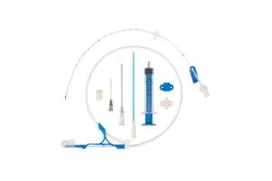
The tip portion of a CVC (Central Venous Catheter) catheter plays a crucial role in its functionality and safety. One important characteristic of the catheter tip is its contrast effect, which refers to the radiopacity or radiodensity of the tip material when visualized using imaging techniques such as X-rays. This contrast effect is essential for accurate placement verification, monitoring, and troubleshooting of the catheter.
In radiology, the contrast effect allows healthcare professionals to precisely determine the position of the catheter tip within the patient’s body. When the catheter is inserted, an X-ray is commonly performed to confirm its placement. The contrast effect enables the tip to be clearly visible on the X-ray image, allowing for accurate localization within the vascular system. This verification step is vital to ensure that the catheter is appropriately positioned in the desired vessel, such as the superior vena cava or one of its tributaries.
Accurate placement of the catheter tip is crucial for several reasons. First, it ensures that medications, fluids, or blood products infused through the catheter are delivered to the intended site. Second, it helps prevent complications associated with malpositioning, such as inadvertent puncture of nearby structures or inadequate flow rates. The contrast effect of the catheter tip aids in achieving precise and reliable placement, reducing the risk of complications and improving patient outcomes.
The contrast effect can also assist in monitoring the catheter during its use. Regular imaging, such as periodic X-rays, allows healthcare providers to assess the catheter’s position and detect any potential migration or malpositioning. This is particularly important when the catheter remains in place for an extended period. By visualizing the contrast effect of the tip, healthcare professionals can promptly identify any changes in position that may require intervention or repositioning of the catheter.
Furthermore, the contrast effect of the catheter tip is valuable in troubleshooting and addressing complications that may arise during catheterization. For example, if a patient experiences symptoms such as catheter malfunction, infection, or thrombosis, imaging can be performed to assess the catheter tip’s position and evaluate potential causes. The contrast effect facilitates the identification of complications, allowing healthcare providers to make informed decisions regarding further management or necessary interventions.
To achieve the contrast effect, different materials and techniques are used in the construction of catheter tips. Radiopaque materials, such as barium sulfate or tungsten, are commonly incorporated into the tip design. These materials have high X-ray attenuation properties, meaning they absorb X-rays more effectively and appear as dense, white areas on the radiographic image. This contrast effect enhances the visibility of the catheter tip against the surrounding tissues and fluids, facilitating accurate localization and assessment.
In addition to the catheter tip itself, the contrast effect can be enhanced by utilizing contrast agents during imaging procedures. Contrast agents are substances that are introduced into the vascular system to improve the visualization of blood vessels and catheter tips. These agents are often iodine-based and can be injected through the catheter to enhance the contrast effect during imaging studies like angiography or computed tomography (CT).
In conclusion, the contrast effect of the tip portion of a CVC catheter is a critical characteristic that aids in accurate placement verification, monitoring, and troubleshooting. By ensuring precise localization of the catheter tip within the patient’s body, healthcare professionals can administer treatments effectively, minimize complications, and provide optimal care. The use of radiopaque materials and contrast agents contributes to achieving the necessary contrast effect, allowing for clear visualization and assessment of the catheter tip during imaging procedures.
